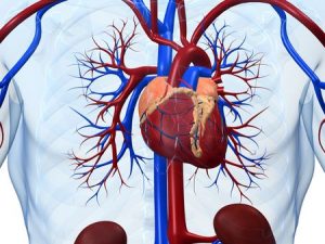
Anatomy of the Heart
As one of the most essential parts of the body, the anatomy of the heart is important to know and understand. The heart is located just behind the sternum, slightly to the left. It’s protected by a tough sac called the pericardium. The heart needs all the protection it can get; it beats an average of 100,000 times a day, pumping about 2,000 gallons worth of blood through the body. The pericardium protects the roots of the major blood vessels. It’s attached to the spinal column and diaphragm with strong ligaments that keep the heart in place and protect it from movement within the chest.
At MIBHS, we are well educated in the anatomy of the heart and want to teach others about it as well. By understanding more about the muscle itself, you can further understand the need to keep it healthy. It may even be a quick recap on the education you already have from growing up in science class. If that is the case, it is never a bad time to refresh your knowledge on parts of the body.
The Anatomy of the Heart Broken Down
The anatomy of the heart is simple once broken into parts. By dividing the heart into its individual sections, it can give patients a greater understanding of the organ itself. This mainly comes down to the walls of the heart, four chambers, and four valves. It may seem impossible to fit that many parts into one organ, but that is part of what makes it such a miraculous part of the body.
Walls of the Heart
The heart’s walls are made up of three main layers; the epicardium, myocardium, and endocardium. Each of these walls have its individual role to ensure your heart is functioning correctly. The epicardium is the outermost layer, a membrane that covers and protects the heart, producing lubricating fluid. The myocardium is what is commonly referred to as the heart’s “muscle.” It is the tissue that contracts and relaxes to produce the heartbeat, pushing the blood through the body. The endocardium is a very smooth layer of tissue that lines the interior of the heart and prevents the formation of blood clots.
4 Chambers
The heart contains four chambers- the left and right atriums, and the left and right ventricles. The atria  make up the upper part of the heart. They are smaller than the ventricles and act as the receiving chamber for blood coming back into the heart, while the ventricles push blood out into the body. There are two circulatory loops attached to the heart. The right loop circulates blood to the lungs, while the left loop pushes oxygenated blood out into the body.
4 Valves
Moving blood through the heart requires four strong valves, one between each of the four chambers and in the arteries and veins that carry blood to and from the heart. The valves control the flow of blood around the heart and ensure the pumping action is efficient. Valves can be replaced surgically using artificial or organic replacements. Taking good care of your heart means enjoying good health throughout your lifetime.
The anatomy of the heart is something that is important for anyone to know. You may even learn more than ever before with this simple read. Take a quick trip back to science class and come away with knowledge about your body and the anatomy of the heart. With walls, valves, and chambers, the organ continues to do its job to keep you healthy and happy. Are you looking to learn more about your heart health? MIBHS is here to help. Check out our website or give us a call for more information.

